- Visibility 82 Views
- Downloads 13 Downloads
- DOI 10.18231/j.jsas.2020.022
-
CrossMark
- Citation
Filariasis presenting as isolated asymptomatic unilateral inguinal lymphadenopathy
- Author Details:
-
Benny PB *
-
Muhsin VK
What’s Known
Filariasis as a rare cause of isolated lymph node enlargement has been described. Diagnosis was established by excision biopsy following imaging studies.
What’s New
Filariasis presenting as isolated asymptomatic unilateral inguinal lymph node enlargement and unilateral hydrosalpinx is rare. Diagnosis via ultrasound imaging, routine blood analysis, excision biopsy and subsequent treatment with diethylcarbamazine resulting in full resolution has not been reported before.
Introduction
Filariasis is a vector borne disease especially prevalent in tropical and subtropical regions and caused by slender thread-like filarial worms from the superfamily of Filarioidea, which has an affinity towards skin and subcutaneous tissue or lymphatic system. While the nematode-like Onchocerca volvulus and Loa loa cause filariasis of the skin, Wuchereria bancrofti, Brugia malayi and Brugia timori cause lymphatic filariasis in the descending order. Wuchereria bancroftian filariasis produces a wide range of clinical manifestations depending upon the phase and duration. The chronic phase is usually characterized by presence of lymphadenopathy in lower limbs, retroperitoneal tissues, lymphedema, hydrocele and elephantiasis. Microfilaria are usually absent in early and late phase of the disease. Circulating filarial antigen testing is more sensitive and can detect the presence of the nematode, even in early stages.
Case Report
A 3-year-old girl was seen at our outpatient clinic with a 1-week history of enlarged painless left inguinal lymphadenopathy. The mass had never been erythematous, and she had never displayed any signs of a systemic infection, including fever or chills. She had never travelled outside the Kollam district. On examination, a single 1.5 × 1.5 cm, mobile, non-tender left inguinal mass with firm consistency was palpated. The rest of the physical examination was unremarkable. Blood routines showed eosinophilia and peripheral smear report came as normocytic normochromic picture.
USG Abdomen and pelvis along with inguinal region revealed left inguinal swelling corresponds to an enlarged lymph node and right sided hydrosalpinx.
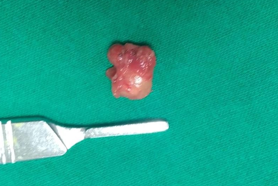
She underwent an excisional biopsy of the left inguinal lymph node, which she tolerated without any complications.
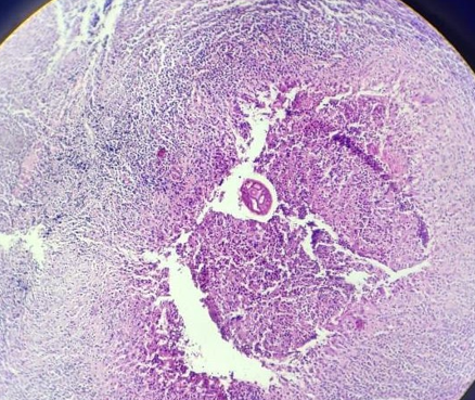
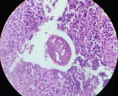
Histopathologic examination revealed cut section of filarial worm surrounded by eosinophilic abscess and marked fibrosis.
She was treated with complete course of Diethylcarbamazine and did a follow up USG of abdomen, which showed complete resolution of right sided hydrosalpinx.


Discussion
Filariae of animals can infect humans and typically produce cryptic infections. These “zoonotic” infections can occur anywhere even non-endemic regions. Infections may be symptomatic or not, and the parasites are found in surgical tissue biopsy specimens or rarely are removed intact from superficial sites such as conjunctiva. Worms have been found in subcutaneous tissues, the heart and lungs, lymphatics, the eye, and the central nervous system.3 Many kinds of filariae have been isolated from humans, including species of Dirofilaria, Brugia, Onchocerca, Dipetalonema, Loaina and Meningonema.[1]
Acute lymphadenitis, asymptomatic subcutaneous swelling, acute lymphangitis, lymph node swellings, funiculoepididymoorchitis, nodules in breast, chronic lymphedema, hydrocele, thyroid or salivary gland, tropical pulmonary eosinophilia and chyluria are the various clinical pictures of lymphatic filariasis.[2] Lymphatic filariasis occurs in nearly 25-30% of all total cases of filariasis while lymph node involvement is found in about 0.047% cases.[2], [3]
Imaging modalities like ultrasonography and magnetic resonance may demonstrate regular twirling motion of echogenic particles of either adult filarial worms or microfilaria in lymphatics (filarial dance sign).[4]
Immunochromatographic and deoxyribonucleic acid probes can demonstrate even species of worm also. This is sensitive and specific method. Fine Needle Aspiration Cytology (FNAC) provides a simple, inexpensive and rapid way to diagnose microfilariae and adult worms. However, finding of microfilaria in FNAC is an uncommon phenomenon.[5]
Superficial sites like lymph nodes, breast lumps, scrotal swellings, thyroid swellings, and soft tissue swellings have very low chances of microfilaria detection.[1], [6], [7], [4], [8], [9], [10] Only 0.078% of detection rate of microfilaria is noted in all samples of lymph node swellings.[2], [11]
Treatment is done by diethylcarbamazine, 6 mg/kg/day for a period of 12 days in divided doses.[8] A single annual dose of oral Albendazole 400 mg and diethylcarbamazine 6 mg/ kg of body weight is recommended for prevention of filariasis in endemic countries.[5]
Vaccines are not effective. Vector control is the key in prevention of transmission.
Conclusion
Lymphatic filariasis, albeit rare, should be considered as a differential diagnosis in lymph node swellings. Ultrasound imaging and Fine-needle aspiration cytology may serve as a useful screening tool in differentiating neoplastic and inflammatory lesions. Excision biopsy is confirmatory. Medical therapy with DEC can produce good results in the filarial lymphadenopathy.
Conflict of Interest
None declared
Source of Funding
None.
References
- TC Orihel, M L Eberhard. Zoonotic Filariasis. Clin Microbiol Rev 1998. [Google Scholar] [Crossref]
- P Gupta, M Bharadwaj, CK Durga. Isolated Epitrochlear Filarial Lymphadenopathy: Cytomorphological Diagnosis of an Unusual Presentation. Turk Patoloji Derg 2020. [Google Scholar] [Crossref]
- S Sivakumar. Role of fine needle aspiration cytology in detection of microfilariae: report of 2 cases. Acta Cytol 2007. [Google Scholar] [Crossref]
- C F Dietrich, N Chaubal, A Hoerauf, K Kling, M S Piontek, L Steffgen. Review of Dancing Parasites in Lymphatic Filariasis. Ultrasound Int Open 2019. [Google Scholar] [Crossref]
- R Raveendran. Lymph Node Enlargement in Neck Filariasis as a Rare Cause: A Case Report. Iran J Med Sci 2017. [Google Scholar]
- . Isolated Epitrochlear Filarial Lymphadenopathy: Cytomorphological Diagnosis of an Unusual Presentation. Turk Patoloji Derg 2018. [Google Scholar] [Crossref]
- DW Kimberlin, MT Brady, MA Jackson, SS Long. American Academy of Pediatrics. Lymphatic Filariasis. 2018 Report of the Committee on Infectious Diseases. American Academy of Pediatrics 2018. [Google Scholar]
- JC Simmonds, M K Mansour, W I Dagher. Cervical Lymphatic Filariasis in a Pediatric Patient: Case Report and Database Analysis of Lymphatic Filariasis in the United States. Am J Trop Med Hygiene 2018. [Google Scholar] [Crossref]
- AS Oguntola, AM Oguntola, OO Bajowa, D Sabageh. Incidental detection of microfilariae in a lymph node aspirate: A case report. Niger Med J 2014. [Google Scholar] [Crossref]
- P T S Kandalam, A N Parampath, H I Farghaly, S A Salah, M K Kayakkool, J V Mathew. An unusual presentation of filariasis in a nonendemic country. Qatar Med J 2015. [Google Scholar] [Crossref]
- P. Pandey, A. Dixit, S. Chandra, A. Tanwar. Cytological diagnosis of bancroftian filariasis presented as a subcutaneous swelling in the cubital fossa: an unusual presentation. Oxford Med Case Rep 2015. [Google Scholar] [Crossref]
How to Cite This Article
Vancouver
PB B, VK M. Filariasis presenting as isolated asymptomatic unilateral inguinal lymphadenopathy [Internet]. IP J Surg Allied Sci. 2025 [cited 2025 Sep 08];2(4):123-125. Available from: https://doi.org/10.18231/j.jsas.2020.022
APA
PB, B., VK, M. (2025). Filariasis presenting as isolated asymptomatic unilateral inguinal lymphadenopathy. IP J Surg Allied Sci, 2(4), 123-125. https://doi.org/10.18231/j.jsas.2020.022
MLA
PB, Benny, VK, Muhsin. "Filariasis presenting as isolated asymptomatic unilateral inguinal lymphadenopathy." IP J Surg Allied Sci, vol. 2, no. 4, 2025, pp. 123-125. https://doi.org/10.18231/j.jsas.2020.022
Chicago
PB, B., VK, M.. "Filariasis presenting as isolated asymptomatic unilateral inguinal lymphadenopathy." IP J Surg Allied Sci 2, no. 4 (2025): 123-125. https://doi.org/10.18231/j.jsas.2020.022
