Introduction
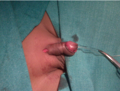
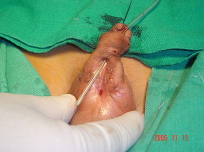
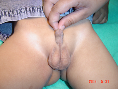
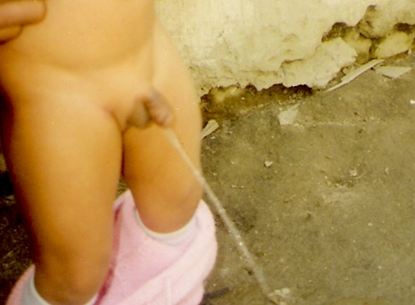
Occurring due to a defective developmental process during embryonic differentiation, this congenital condition often raises a doubt regarding the patients’ gender. This is more so in the more severe of the cases. Striking is the “Monk appearance” due to the “hooded” prepuce, deficient ventrally, although the main complaint of the parents might be a thin urine stream from an abnormal site on the ventral side of the penis. Hundreds of techniques are described in literature for Hypospadius, which in themselves attest to the fact that no single technique is the perfect solution to this complex problem. Starting from the Dennis Browne technique to the authors’ recently claiming near perfect corrections, there is a big gap between the patient numbers and the exponents available to perform this surgery. This is the reason that the authors wish to enumerate upon a generilised principles to steer surgeons and beginners through almost types of cases rather than stress upon techniques.
Aims and Objectives
The presenting author is a student of Dr. H.S. Asopa and an advocate of the prepucial island flap technique, though not dogmatic in its propagation. He has also had the opportunity to train with Mr. Aviar Bracka and uses different techniques judiciously.1
This presentation though is to give a step by step detail of how to go about the surgery for Hypospadius and what to avoid. The author does not advocate a particular technique or stage/single stage method but will be stressing upon the finer points comparing different methods and connected outcomes.
Materials and Methods
The authors have undertaken about 3600 fresh cases of hypospadius repairs spaced over almost two decades. This is also a journey of the author’s experience over the years of this challenging condition. The authors have been performing single stage as well as two staged repairs. Not included in the count are cases which were previously termed as “Hypospadius cripple” although treated with the same techniques with similar outcomes.
The single stage repairs were done mostly by the Asopa-1 (Jalandhar modification) technique performing a patch or patch tube patch urethroplasty. The multi staged repairs were done by the Bracka technique using the buccal mucosa as the graft for the urethral plate.2
In addition there were also cases of MAGPI and flip flap performed for glandular and coronal hypospadius respectively.
Discussion
The answers to correction of this challenging deformity probably lie in discussing each issue concerning the surgery and not just the technique itself. Some of the common problems in Hypospadius surgery which need to be addressed to achieve consistently satisfactory results are as follows:-
The ten commandments of hypospadius surgery
Who should perform it and when? This answer is very simple. The surgeon who is best trained to undertake this procedure. There has always been a conflict amongst the different specialists on this issue. We are of the opinion that out of the plastic surgeon, urologist, pediatric surgeon or general surgeon, the best suited is the plastic surgeon as he/she is adept in tissue handling. But the significant factor is the training and experience. It is best to perform this surgery before the age of 4 years, which is the age of recall. Children operated after this have a memory of the surgery which could lead to psychological problems.3
The interaction of the surgeon and the guardians is extremely important in the post operative course. Parents who do not follow the instructions of the doctor and staff often see poor results in their wards. In centers where the nursing care is good and parents are not given charge of the patients of the first few days end up with consistently good results.
Which technique is to be used is a very difficult question to answer, as well the simplest. Each surgeon over the years starts performing that technique with which he/she keeps getting consistently good results. As mentioned earlier, no technique is perfect. There are hundreds of described techniques for this surgery, which is a proof that none is the perfect answer. It is only the skill and experience of the surgeon which is important.4
Single v/s two staged. The concept of staging has undergone a drastic change over the years. Historically the concept of staging was based on the thought that chordee needed to be removed completely at stage one. The surgeons also undertook the liberty to transpose the prepuce ventrally to make up for the tissue deficiency. Chordee was also considered to be a growing tissue requiring repeated surgeries.Over time we realized that chordee is congenital and can be removed in one surgery. The transposition of dorsal hood to the ventrum was substituted by the use of perputial tube or patch urethroplasty.The current concept of staged repair is confined to cases which need a health urethral plate on which to build the neo-urethra. In cases lacking a health urethral plate, buccal or bladder mucosa is used to create a health urethral plate and also excising any chordee which may persist. The second stage is for the definitive urethroplasty. (Mr. Snodgrass prefers to perform this in a single stage).
What anesthesia should be used? All young patients should be done in GA. We have also used caudal block, which is also useful for post operative analgesia. Ketamine or spinal anesthesia should be avoided in adults as it could cause a troublesome erection.
Use of tourniquet/ infiltration. We regularly use infiltration with xylocaine2% with adrenaline diluted in the ration of 1:4. The liberal use of this is sufficient to provide a bloodless field for repair, if the surgeon displays patience after the infiltration. Tourniquet is useful for the artificial erection test at the start of surgery or after correction of the chordee. The use of tourniquet during the procedure has been abandoned as cumbersome and could even be harmful to the patient. 5
Prevention of post operative edema. Edema is the bugbear of all surgeries. The penis being like a box canyon (dead end) is prone to post operative edema. The ensuring of proper haemostasis, use of good quality dressing providing uniform pressure and use of drain, if in doubt, prevent edema in most cases. The use of a dorsal slit incision at the root of the penis prevents the band effect which is responsible for edema in most cases. Surgeons are advised to use this in all cases.
A good surgical repair is the best for preventing fistulae. Using a dartos waterproofing layer over the inner repair provides a protective layer between the two suture lines which should ideally not overlap. Dr.Asopa advocated the use of inverted sutures for the inner repair as well as use of subcutaneous sutures for the skin.1 This goes a long way in preventing fistulae. Indwelling catheter is used for 14 days in most penile or proximal repairs. A voiding trial is done at the time of removal of catheter. In case some small fistula is seen, the catheter can be used for another 10 days. Any persistent fistula should not be attempted before a 3 month post operative period.6
Numerous late complications can appear after a long term follow up. Strictures may occur at the neo-meatus or the junction of the neo-urethra with the main urethra. Strictures seldom respond to dilatation. There can be diverticulum formation which needs surgical correction (usually associated with a stricture). There can also be hair growth in the neo urethra in case the prepuce is used for repair. Most of these need to be dealt with definitely with surgical intervention.7
The question of aesthetic appearance has gained importance in the recent years as the opposite sex has become very demanding in this respect and a poor aesthetic appearance can lead to serious psychological problems in the patient. It is for this reason that techniques like Snodgrass and Bracka are becoming popular as they provide a good aesthetic result.8
Tissue handling is single most imp step in achieving consistently good results. It is often stressed upon that delicate handling of tissues is very important, more so in cases of such deformities. The example to compare is of a cat picking up its kittens. Firmly, but without any damage due to its teeth on the subject.9
Results
Over the years we have been achieving consistent results even though we have not struck to any one technique. We have been using the modified Asopa-I technique (chosen mostly for patients who are poor, uneducated and for social reasons might not be able to come for or afford a second surgery) and Bracka technique (where the aesthetic appearance and outcome are as important to the patient and family as the functional results) as the two main methods for surgery.2 This step by step on discussion in hypospadius surgical repair is not to highlight surgical techniques but to discuss about steps which have their own importance to produce acceptable results.10
Conclusion
In spite of so many methods to perform surgery for patients of hypospadius, some general principles on tissue handling, suture management, prevention of edema, general methods of dissection, dressing techniques etc go a long way in giving consistent results whatever the method used for the repair.11, 12


