Introduction
The formation of brachial plexus is complex, as it is formed by the union of ventral rami of C 5 6 7 & 8 and the T1 with a twig from C4 and T2. These rami unite to form the trunks of brachial plexus namely upper middle and lower trunks, each trunk divides into divisions namely anterior and posterior divisions out of which all posterior divisions unite to form the posterior cord. The Lateral cord is formed by the union of anterior divisions of upper and middle trunks of brachial plexus. These cords and their branches appear in the axilla grouped around the axillary artery. The lateral cord gives its first branch namely the lateral pectoral nerve to the pectoralis major muscle and then divides into musculocutaneous and lateral root of median nerve.
The Musculocutaneous Nerve classically pierces the coracobrachialis muscle and passes obliquely to the lateral side of the arm between the biceps brachii and the brachialis muscle, also supplies both of them. Later it pierces the deep facia above elbow lateral to the tendon of the biceps brachii and continues as the lateral cutaneous nerve of the forearm.
The lateral root of median nerve is the largest branch of the lateral cord of brachial plexus, joins the medial root coming from the medial cord and forms the Median nerve in front of the Axillary artery in the Axilla.
The variations of the lateral cord of brachial plexus should be kept in mind while performing surgical exploration of the axilla, Shoulder region and arm region to avoid damage to these important nerves.
Materials and Methods
The study was carried out to determine the branches of the lateral cord of the brachial plexus in the region of axilla. The specimens were neatly dissected out before giving it to the undergraduate students for routine dissection in the Anatomy dissection hall. A total of 50 limbs were selected for the present study irrespective of the gender. These 50 specimens were collected from 10% formalin fixed human cadavers, no special mode of preservation was required for the purpose of the study and was done during routine undergraduate dissection in the Department of Anatomy, over a period of 5 years by routine procedure of standard dissection of cadavers. 20 already dissected specimens were also used if structures were not disturbed. Thus both New and old dissected limbs were used for the purpose of study Detailed dissection was carried out in the limbs in the region of Axilla and the branches were traced till the end.
Observations
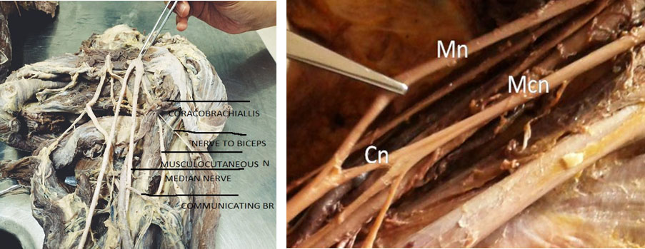
Out of the 50 limbs selected for the present study, three limbs showed the musculo-cutaneous nerve coming from the lateral root of the median nerve instead of the lateral cord of brachial plexus.
Out of the three originating from the lateral root of median and only one was bilateral and the rest were seen on left side only with normal origin on the Right side.
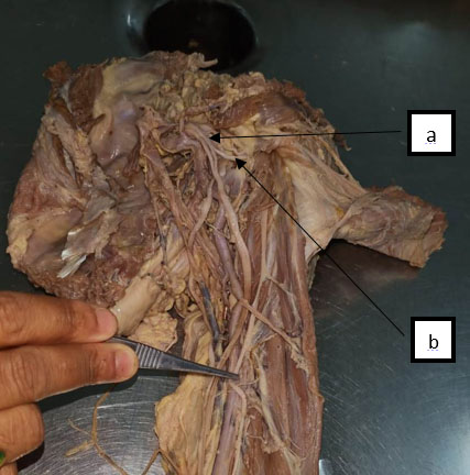
Level of formation of Lateral cord was noted in every specimen, none of the specimens showed higher level of formation but one specimen showed divisions of lateral cord into its branches at a lower level compared to the other side but no signigicant variations found in the branching pattern of the lateral cord was seen in that specimen.
In one specimen Musculocutaneous nerve was seen not piercing the Coracobrachialis, but giving a branch to it instead.
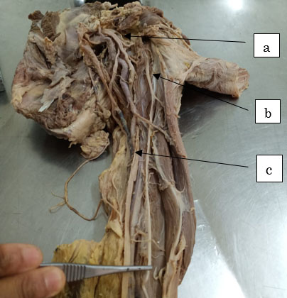
In one Musculocutaneous nerve was absent and all the muscles were supplied by the branches from the Median nerve.
In one specimen the Musculocutaneous nerve ends by supplying the Coraco brachialis and the rest of the muscles were supplied by branches from Median nerve. The lateral cutaneous nerve of forearm which should be the continuation of Musculocutaneous nerve was a branch from Median nerve.
Discussion
Coracobrachialis being one of the flexor muscles of anterior compartment of arm along with the biceps and brachialis muscles is highly variable muscle as it is reported having 2 bellies which itself is a topic of research but when a variation of coracobrachialis is observed the nerve supplying it being the musculocutaneous nerve a variation in it is also expected.
The musculocutaneous nerve as such is known for multiple variations in its branching pattern while supplying the muscles of anterior compartment of arm before terminating as lateral cutaneous nerve of the forearm.
Anomalies in the formation of lateral cord of brachial plexus and the communication between its branches are being observed commonly but the variation of the course of lateral cord is not much reported in the literature.1
Variations of the median and musculocutaneous nerve at the level of brachial plexus are common. Anastomosis between the MCN and the MN is by far the most common and frequent of all the variations that are observed among the branches of the brachial plexus (Venieratos and Anangnostopoulou, 1998).2
The variations in coracobrachialis and the variations of branching pattern of the musculocutaneous nerve are taken up for further research in due course of time giving preference to the occupation as coracobrachialis is variable morphologically and depending on the occupation hence on wide range of population needs to be the target.
Coracobrachialis is a flexor muscle of the arm and is vulnerable to the injury from the retractors placed under the coracoid muscles as required during shoulder reconstructive surgery.
The operative management by coracoid graft transfers in the recurrent dislocations of shoulder and shoulder arthroscopies could be the source of lesions to the structures piercing the muscle. (Flatow et al, 1989; Laburthe-Tolra, 1994-95).3 The muscle has been suggested for possible use as flap for coverage in infraclavicular defects of exposed axillary vessels, especially in postmastecomy reconstructive surgery (Hober et al, 1990).4
The interpretation of the anomaly of the atypical course of lateral cord requires consideration of the development and innervation of upper limb musculature. Muscles of the limbs are derived fromsomatic precursor muscle cells from theventrolateral edges of the somites opposite thedeveloping limbs, which lie lateral to the neural tubeand causes bulge in the overlying ectoderm.Somites have a specific effect on the position of thedeveloping spinal nerves, which preferentially growthrough the cranial half of sclerotome. Spinal nervesare derived from two sources, the motor nerve fromthe neural tube and the sensory nerves from the neural crest (Williams et al, 1995). The nerve cordsfrom the spinal nerves that correspond to the early extent of limb buds grow distally to establish anintimate contact with the differentiating mesodermal condensations into intermuscular spaces and end ina-premuscle mass.
As suggested by Sannes et al(2000) that the guidance of the developing axons is regulated by expression of chemoattractants and chemorepulsants in a highly coordinated sitespecific fashion. Any alterations in signaling between mesenchymal cells and neuronal growthcones can lead to significant variations and probablyin this case causes the lateral cord to pass throughthe coracobrachialis muscle. Once formed,anydevelopmental differences would persist postnatally. (Brown et al. 1991;2 Arey, 1966).5
The study can help us analysing the Neuropathy followed by trauma or injury to the brachial plexus leading to the involvement of musculocutaneous nerve as a singled out sign or symptom The injury to brachial plexus though common during accidental fall or injuries isolated injuries to musculocutaneous nerve or lateral cord in perticular is rare but can be sole symptom from patient with history of fall or accident in those cases the knowledge of the variations in the branching pattern may be of help to relive the patient with probable neurological symptoms he may be facing. Lower the division of lateral cord higher are the chances of variation in the branches which again is a topic for further research.6, 7, 8, 9, 10
The knowledge of the course and distribution of the lateral cord of brachial plexus, keeping in mind the variations in anatomy and the level of penetration are important while performing shoulder arthroscopy by anterior glenohumeral portal and shoulder reconstructive surgeries.


